Thursday 28 August 2014

Origin/Insertion of Muscles - Abductor Muscles (Gluteus Maximus, Gluteus Medius)

The hip abductor muscles are a group of 4 muscles, Gluteus Maximus, Gluteus Medius, Gluteus Minimus and Tensor Fascia Latae, that are located in the butt. Their main function is to abduct (or separate the legs) away from the midline of the body. It occurs in any athletic movement requiring side-to-side movement.

Gluteus Maximus
Origin: Surface of Ilium posterior to posterior gluteal line and posterior inferior surface of sacrum and coccyx
Insertion: Gluteal tuberosity of femur; anterior aspect of lateral tibial condyle via the Iliotibial tract (band)
Action: Extends, laterally rotates thigh at hip; upper fibres help to abduct the thigh (the inferior fibres also stabilise the extended knee by tightening the Iliotibial tract)

Gluteus Maximus


Gluteus Maximus Origin


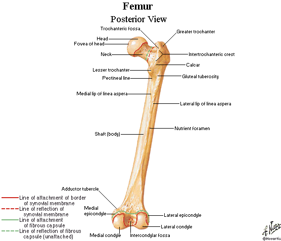
Gluteal Tuberosity
Gluteus Medius
Origin: Outer surface of Ilium, between the posterior and anterior gluteal lines
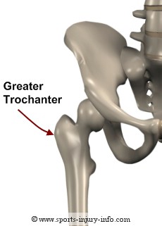
Insertion: Lateral and superior surfaces of the greater trochanter of femur
Action: Abducts thigh at hip; anterior fibres medially rotate thigh and posterior fibres laterally rotate thigh at hip

Gluteus Medius

Gluteal Lines

Greater Trochanter


Wednesday 20 August 2014

Origin/Insertion of Muscles - Biceps, Brachialis and Brachioradialis

I wasn't original going to do the origin and insertion of these muscles now. However, I am putting it in here because I have a project to do for work. Which I will tell you about at a later date. It involves a video and I am a little anxious about it as I am still learning all this!!!!!!! But is there any other way to learn, other than getting in there and just DOING it!
 
Biceps Brachii, Brachialis and Brachioradialis
 
Biceps Brachii
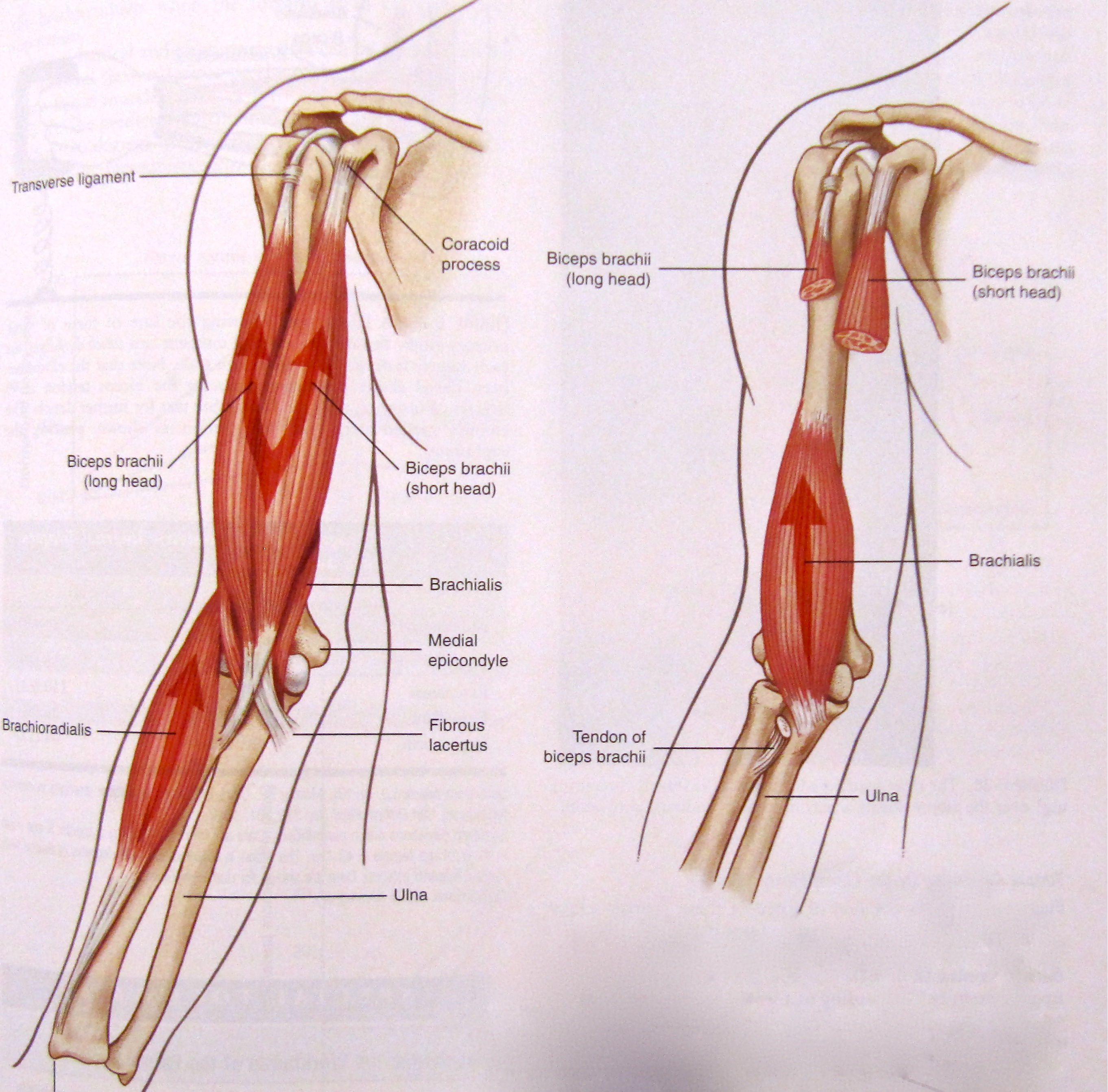
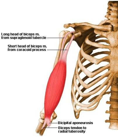
Is a two headed muscles that crosses both the shoulder and elbow joints. It is one of 3 muscles that flex the forearm at the elbow.

Origin: Long Head: Supraglenoid tubercle of the scapula
            Short Head: Coracoid process of the scapula
Insertion: Radial tuberosity; bicipital aponeurosis to the fascia on the medial side of the forearm
Action: Flexes the forearm at the elbow; also flexes arm (weakly) at the shoulder

Supraglenoid Tubercle and Coracoid Process
 
Radial Tuberosity

Bicipital aponeurosis is "a flat sheet of connective tissue that fans out from the medial side of the tendon to blend with deep fascia covering the anterior compartment of the forearm". http://medical-dictionary.thefreedictionary.com/Lacertus+fibrosus

Brachialis
Origin: Anterior, distal half of humerus
Insertion: Coracoid process and tuberosity of ulna
Action: Flexes forearm at elbow

Anterior, distal half of humerus

Coracoid process of ulna

Brachioradialis
Origin: Lateral supracondylar ridge of humerus
Insertion: Styloid process of radius
Action: Flexes the arm at elbow

Lateral Supracondylar ridge
 
Styloid Process

Monday 18 August 2014

Origin/Insertion of Muscles - Adductor Muscles (Gracilis and Pectineus)

Gracilis
Origin: Inferior border of pubic body near pubic symphysis
Insertion: Proximal medial surface of tibia
Action: Adducts thigh at hip; flexes leg at knee and medially rotates leg when knee is flexed


Gracilis Muscle
 

Origin: Inferior Pubic Ramus
Insertion: Proximal tibia, medially
Pectineus
Origin: Pectineal line along superior ramus of pubis
Insertion: Posterior surface of femur, inferior to the lesser trochanter
Action: Adducts and flexes thigh at hip
 
Pectineus Muscle
 
Pectineal Line of Superior ramus of Pubis
 
Insertion: Posterior surface of femur; inferior to lesser trochanter
 

Wednesday 13 August 2014

Setting Goals

Saw this clip tonight and thought it would be interesting for you to watch. I know it will come in handy for me when setting goals with my Personal Training clients.

https://www.youtube.com/watch?v=sk8hNqkhXD8&feature=youtu.be

Joce

Origin/Insertion of Muscles - Adductor Muscles (Adductor Brevis, Longus and Magus)

The adductor muscles work to pull your leg to the midline of the body. These muscles include:
  • Adductor Brevis
  • Adductor Longus
  • Adductor Magus
  • Adductor Minimus
  • Pectineus
  • Gracilis
The Adductors Brevis, Longus and Magus and Gracilis are the hip joint adductors.

Adductor Muscles


Adductor Brevis
Origin: Lateral (outer) surface of the inferior ramus of pubis
Insertion: Proximal portion of linea aspera
Actions: Adducts and flexes thigh at hip

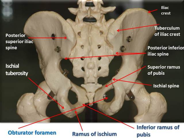
Inferior Pubic Ramus


Linea Aspera
 
 
Adductor Longus
Origin: Superior aspect of pubis, below pubic tubercle
Insertion: Middle third of linea aspera of femur along medial hip
Action: Adducts and flexes thigh at hip


Superior Ramus of Pubis
(Great labelling for all the adductor muscles listed in this post)

Adductor Longus

 
Adductor Magus
Origin: Oblique Head: Inferior ramus of pubis and ischial ramus
             Vertical Head: Ischial Tuberosity
Insertion: Oblique Head: Gluteal Tuberosity, linea aspera and proximal  supracondylar line of femur
                Vertical Head: Adductor tubercle of femur
Action: Oblique: Adducts and flexes thigh at hip
             Vertical: Extends thigh at hip
 
Ischial Ramus
Ischial Tuberosity


Gluteal Tuberosity and Linea Aspera

Supracondylar Line

Adductor Tubercle of Femur

Monday 11 August 2014

Terminology

There is a lot of terminology being thrown around. Words like proximal, distal, medial and lateral. So lets take a look at what all this terminology means!!

Planes of Body Motion
Sagittal: Plane passes through the body from anterior to posterior. It divides the body into left and right
Frontal: Plane passes through the body from lateral to medial. It divides the body into front (anterior) and back (posterior)
Transverse: Plane passes through the body level with waist. It divides the body into upper (superior) and lower (inferior)

Directional Terminology
Superior: Towards to head
Inferior: Towards the feet
Anterior: Toward the front of the body
Posterior: Toward the back of the body
Medial: Toward the midline of the body
Lateral: Toward the side of the body
Proximal: Towards the attachment point of a limb
Distal: Away from the attachment point of a limb

Movement Terminology
Flexion: To decrease the angle of a joint
Extension: Increases the angle of a joint
Abduction: Increase in angle of a limb away from body
Adduction: Decrease of angle of limb away from body (to remember this I usually find myself saying if I add something I am adding it to the body)
Lateral/External rotation: Rotate limb outwards, around long axis of bone
Medial/Internal rotation: Rotate limb inwards, around long axis of bone
Protraction: Bring body part forwards. Protrude a body part (jaw, shoulder blade)
Retraction: Bring body part backwards
Elevation: Lift a body part
Depression: Lower a body part
Supination: Turn palm anterior
Pronation: Turn palm posterior
Inversion: Turn sole of foot medially
Eversion: Turn sole of foot laterally
Dorsiflexion: Bring toes towards ankle
Plantarflexion: Bring toes toward the floor

From - The Essential Guide to Fitness - Rosemary Marchese and Andrew Hill

Sunday 10 August 2014

Origin/Insertion of Muscles - Lower Leg - Gastrocnemius and Soleus

Gastrocnemius
Origin: Medial Head: Medial condyle of femur and area just above the condyle
             Lateral Head: Lateral condyle of femur and area just above condyle
Insertion: Posterior calcaneus via calcaneal (Achilles) tendon
Action: Flexes leg at knee; plantar flexes foot


Medial and Lateral Condyle of Femur


Calcaneal (Achilles) Tendon

Soleus
Origin: Proximal half of posterior surface of the tibia along soleal line; proximal 1/3 of posterior fibular
Insertion: Posterior calcaneus via calcaneal (Achilles) tendon
Action: Plantar flexes foot

Soleal Line


Thursday 7 August 2014

Origin/Insertion of Muscles - Hamstrings - Semimembranosus and Semitendinosus

Semimembranosus
Origin: Ischial Tuberosity of os coxa
Insertion: Posterior medial condyle of tibia
Action: Flexes leg at knee, medially rotates leg at knee when knee is flexed; also extends thigh at hip


Ischial Tuberosity

Posterior Medial Condyle (Tibia)
Semitendinosus
Origin: Ischial Tuberosity of os coxa
Insertion: Proximal tibia, medial to the tibial tuberosity
Action: Flexes leg at knee, medially rotates leg at knee when knee is flexed
Proximal Tibia (Posterior view)

Origin/Insertion of Muscles - Hamstrings - Biceps Femoris

The Hamstrings:

There are 3 separate muscles that make up the hamstring group:
  • Biceps Femoris
  • Semimembranosus
  • Semitendinous
The biceps femoris has 2 heads, the long head and short head.

Biceps Femoris (Long Head)
Origin: Ischial tuberosity of os coxa
Insertion: Head of fibula and lateral condyle of tibia
Action: Flexes leg at knee and laterally rotates leg at knee when knee is flexed; also extends thigh at hip

Biceps Femoris (Short Head)
Origin: Distal linea aspera and lateral supracondylar ridge of femur
Insertion: Head of fibula and lateral condyle of tibia
Action: Flexes leg at knee and laterally rotates leg at knee when knee is flexed

  
Ischial Tuberosity
 Lateral Supracondylar Ridge
Lateral Condyle of Tibia


10 Reasons to get fit!


So I saw this on my Facebook page. I had saved it there a long time ago. I thought it was appropriate to put it on here. 10 good reasons why you should start exercising today!! I did 30 minutes of interval training on the treadmill! What did you do?

Joce xx

Tuesday 5 August 2014

Origin/Insertion of Muscles - Sartorius

Sartorius:
Origin: Anterior Superior Iliac Spine (ASIS)
Insertion: Proximal tibia, medial to tibial tuberosity
Action: Flexes, abducts, laterally rotates thigh at hip; also flexes leg at knee


Anterior Superior Iliac Spine (ASIS)

So where is this in relation to the Anterior Inferior Iliac Spine (AIIS), which is the origin of the Rectus Femoris:

Anterior Inferior Iliac Spine
 
Insertion of Sartorius is shown below, the proximal tibia:


Origin/Insertion of Muscles - Quadriceps - Vastus Intermedius

Vastus Intermedius:
Origin: Anterior and lateral surfaces of the shaft of femur
Insertion: Quadriceps tendon to the base of patella and onto the tibial tuberosity via patella tendon
Action: Extends leg at knee


Below is a greater picture of the Vastus Medius, Vastus Lateralis and Vastus Intermedius. Each shows the Origin and Insertion points.


Origin/Insertion of Muscles - Quadriceps - Vastus Lateralis

Vastus Lateralis:
Origin: Greater trochanter. Also attaches to the lip of the lateral lip of the linea aspera of femur
Insertion: Quadriceps tendon to the base of the patella and onto the tibial tuberosity via the patella ligament
Action: Extends leg at knee


Lateral lip of Linea Aspera
Greater Trochanter


Sunday 3 August 2014

Origin/Insertion of Muscles - Quadriceps - Vastus Medialis

Vastus Medialis:
Origin: Intertrochanteric line
Insertion: Quadriceps tendon to the base of the patella and onto the tibial tuberosity via the patellar ligament
Action: Extends the leg at knee


Intertrochanteric Line

Origin/Insertion of Muscles - Quadriceps - Rectus Femoris

The quadriceps are made up of 4 muscles:
  • Rectus Femoris
  • Vastus Medialis
  • Vastus Lateralis
  • Vastus Intermedius
Functions of the quadriceps:
  • Flexes thigh at hip, extends leg at knee, adducts leg
  • Allows the upper leg to move
  • Reacts with the hamstrings and gastrocnemius
  • Keeps leg straight when standing
Rectus Femoris:
Origin: Anterior Inferior Iliac Spine
Insertion: Quadriceps tendon to the base of the patella and onto the tibial tuberosity via the patellar ligament
Action: Flexes thigh at hip, extends leg at knee


Anterior Inferior Iliac Spine

  Tibial Tuberosity

VO2 Max

VO2 Max is the maximum amount of oxygen we can utilise during intense (maximal) exercise. It is measured in millilitres of oxygen per minute per kilogram of body weight. This is one factor that will help determine a persons capacity to perform sustained exercise. It is often linked to aerobic endurance.

There is a test that can be used as an estimate of a persons VO2 Max called the Bruce Treadmill test. You can see this test at the link below:
https://www.youtube.com/watch?v=A2z0l9B6aGE

This test is considered the best indicator of a persons cardiovascular fitness and aerobic endurance. High VO2 Max is an indicator of an athletes aerobic endurance potential. During high intensity exercise the more oxygen you can utilise the more ATP you can produce. The higher a persons VO2 Max the more oxygen they are able to utilise.

VO2 max has a genetic component which can be increased through training. There are 2 methods used to help increase VO2 Max, these are training volume and training intensity. As a beginner you can increase VO2 Max by 20% with the appropriate training. Elite athletes find this much more difficult, potentially due to the fact that they are likely to be nearer to their genetic potential.

There are 3 factors that influence VO2 Max:
  • Age: Generally our VO2 Max is at its highest by age 20. It decreases up to 30% by age 65
  • Gender: Female elite athletes generally have higher VO2 Max levels than man. However, due to the differences in body size, composition, blood volume and haemoglobin content between men and women, a women's VO2 Max is about 20% lover than a mans
  • Altitude: As there is less oxygen at higher altitudes athletes will have a decrease in VO2 Max  results of roughly 5 %  with a 5000 feet gain in altitude